
The modern diagnostic landscape is increasingly reliant on advanced imaging techniques, and the carotid ultrasound report is a cornerstone of this process. It provides a detailed visual representation of the carotid artery, allowing clinicians to assess its structure, blood flow, and potential abnormalities. A well-structured and accurate carotid ultrasound report is crucial for guiding treatment decisions and monitoring patient outcomes. This article will delve into the intricacies of creating a comprehensive and effective Carotid Ultrasound Report Template, covering everything from preparation to interpretation. Understanding the nuances of this report is vital for healthcare professionals seeking to optimize patient care. Carotid Ultrasound Report Template – a standardized format ensures consistency and facilitates efficient communication among specialists. This guide will equip you with the knowledge to produce reports that are both informative and clinically useful.
Understanding the Basics of Carotid Ultrasound
Before we dive into the template itself, it's important to grasp the fundamental principles behind carotid ultrasound. This imaging technique utilizes high-frequency sound waves to create detailed images of the carotid artery. Unlike traditional X-rays, which provide limited visualization, ultrasound offers superior resolution, allowing clinicians to identify subtle abnormalities that might be missed by other methods. The process typically involves a linear transducer placed across the carotid artery, emitting sound waves that bounce off the vessel walls and are detected by a receiver. Different imaging modes, such as Doppler ultrasound, are employed to assess blood flow velocity and identify areas of stenosis (narrowing) or thrombosis (blood clot formation). The report generated is a visual representation of these findings, providing a snapshot of the artery's health.

The Components of a Carotid Ultrasound Report
A complete carotid ultrasound report typically includes several key sections, each addressing a specific aspect of the artery's anatomy and function. These sections are designed to provide a holistic view of the vessel's health. The report is usually formatted in a standardized manner, allowing for easy comparison and analysis across different clinicians. Here's a breakdown of the essential components:
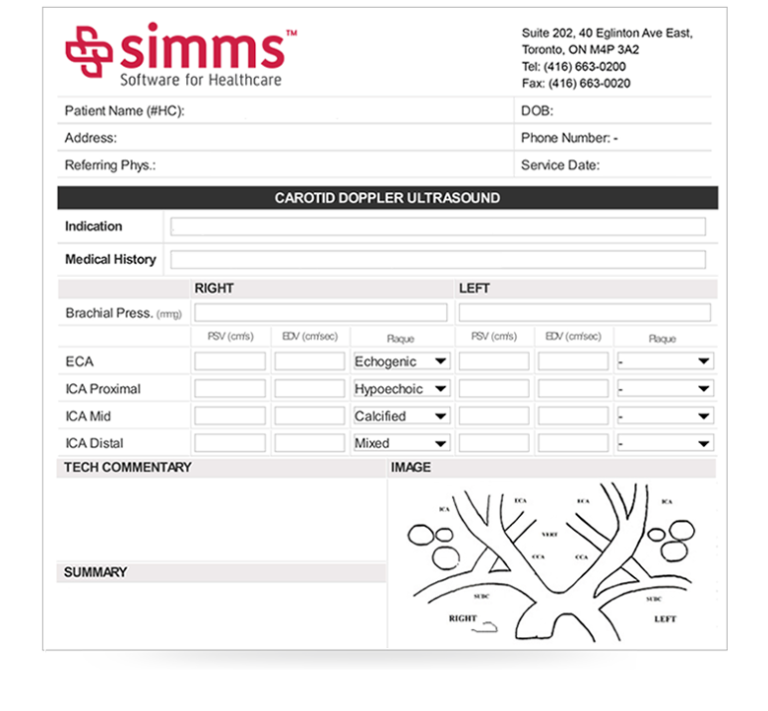
- Patient Identification: The report begins with the patient's unique identifier, ensuring accurate record-keeping.
- Date and Time of Examination: Precise timestamps are crucial for tracking trends and correlating findings with clinical events.
- Imaging Parameters: Details about the ultrasound settings, including transducer type, frequency, and gain, are included.
- Image Acquisition: A description of the image quality, including any artifacts or limitations encountered during the scan.
- Findings: This is the core of the report, detailing the observed characteristics of the carotid artery.
- Assessment: A clinician's interpretation of the findings, including any diagnoses or recommendations.
Section 1: Image Acquisition – A Detailed Overview
The quality of the ultrasound image is directly dependent on the acquisition parameters. Several factors influence the clarity and resolution of the scan, and careful attention to these details is essential. Carotid Ultrasound Report Template relies heavily on precise imaging parameters to ensure accurate representation of the vessel. Common parameters include:

- Frequency: The frequency of the sound waves used. Higher frequencies generally provide better resolution but can be more susceptible to artifacts.
- Gain: The intensity of the ultrasound signal. Adjusting the gain optimizes image clarity while minimizing noise.
- Pulse Width: The duration of the ultrasound pulse. A longer pulse width provides more detail but can increase scan time.
- Translation Speed: The speed at which the transducer moves across the carotid artery. Faster translation speeds improve image sharpness but can be challenging for patients.
It's important to note that variations in these parameters can significantly impact the final image quality. Clinicians should carefully adjust these settings based on the specific patient and the clinical context. Regular calibration of the ultrasound equipment is also vital to maintain optimal performance.
Section 2: Findings – Identifying Abnormalities
This section is where the clinical significance of the ultrasound findings is elucidated. It's crucial to be specific and objective in describing any abnormalities observed. Here are some common findings and how they are typically described:
- Calcifications: The presence of calcium deposits within the artery wall. These can be described as "small calcifications," "large calcifications," or "multiple calcifications."
- Stenosis: Narrowing of the artery, often caused by plaque buildup. This is typically described as "mild stenosis," "moderate stenosis," or "severe stenosis."
- Thrombosis: The presence of blood clots within the artery. This is often identified as "small thrombus," "moderate thrombus," or "large thrombus."
- Aneurysms: Bulges in the artery wall, which can compromise blood flow. These are typically described as "small aneurysm," "moderate aneurysm," or "large aneurysm."
- Branch Abnormalities: Changes in the branching patterns of the artery, which can indicate vascular disease.
- Lymphoid Enhancement: The presence of increased blood flow in the lymphatics surrounding the artery, which can be a sign of inflammation or infection.
It's important to note that the interpretation of findings should always be done in conjunction with the patient's clinical history and other imaging modalities.
Section 3: Assessment – Clinical Implications
This section provides a concise summary of the clinical significance of the findings. It's not just about identifying the abnormality; it's about understanding its potential impact on the patient. For example:
- Small Calcifications: May be associated with increased risk of stroke.
- Moderate Stenosis: Can lead to reduced blood flow and symptoms such as pain, numbness, or tingling.
- Thrombosis: Requires prompt treatment to prevent complications such as stroke or limb ischemia.
- Aneurysms: Require immediate intervention to prevent rupture.
The assessment should consider the patient's symptoms, risk factors, and overall clinical picture.
Section 4: Additional Considerations – Reporting and Documentation
Beyond the core sections, a comprehensive carotid ultrasound report should include additional considerations:
- Image Quality Assessment: A brief assessment of the image quality, noting any artifacts or limitations.
- Patient Demographics: Information about the patient's age, sex, and medical history.
- Ordering Physician: The name of the physician who ordered the ultrasound.
- Date and Time of Report: As mentioned earlier, precise timestamps are crucial.
- Unique Identifier: A unique identifier for the report to facilitate tracking and retrieval.
Conclusion – The Importance of Accurate Reporting
The Carotid Ultrasound Report Template is a vital tool for clinicians, providing a standardized framework for assessing carotid artery health. By adhering to best practices for image acquisition, interpretation, and documentation, healthcare professionals can ensure that patients receive timely and accurate diagnoses, leading to improved patient outcomes. The consistent application of this template contributes significantly to the overall quality of care delivered. Ultimately, a well-executed carotid ultrasound report empowers clinicians to make informed decisions and provide optimal treatment strategies. The continued refinement of this template, incorporating advancements in ultrasound technology and clinical guidelines, will undoubtedly enhance its effectiveness in the years to come. Carotid Ultrasound Report Template – a tool that demands precision and attention to detail.
0 Response to "Carotid Ultrasound Report Template"
Posting Komentar